
Proteomics: Two Molecular Subtypes in Lupus Nephritis
Renal biopsy still decides how lupus nephritis is treated, yet the ISN/RPS class a pathologist assigns says surprisingly little about who will lose kidney function. A group working across four tertiary hospitals took aim at that gap with proteomics, running data-independent acquisition LC-MS/MS on formalin-fixed paraffin-embedded biopsy sections from 292 patients. About 5,000 proteins survived quality filtering. Unsupervised clustering split the cohort into two molecular subtypes that track with renal function and chronic damage rather than with histologic class, and one separating protein, APOL1, was measurable again in serum.
Key Takeaways
Archival FFPE kidney biopsies are workable input for cohort-scale proteomics: 292 lupus nephritis blocks yielded roughly 5,000 quantified proteins.
Lupus nephritis resolves into two reproducible molecular subtypes, one driven by interferon signaling and complement activation, the other by epithelial structural remodeling.
APOL1 separated the subtypes in tissue and again in serum by ELISA, making it a testable blood-based stratification candidate rather than a tissue-only marker.
A five-protein panel told proliferative from membranous disease at an AUC near 0.76, useful for triage but nowhere near a biopsy replacement.
What FFPE Proteomics Adds to the Lupus Nephritis Biopsy
The analytical setup was deliberately plain: peptides from whole biopsy sections onto a nanoLC system coupled to a timsTOF Pro2 in diaPASEF mode, 25 isolation windows across m/z 400 to 1000, a 60-minute gradient, roughly 200 ng on column. That consistency matters more than it sounds. Formalin crosslinking and variable block age make FFPE the least forgiving matrix in mass spectrometry, and uneven peptide load is the fastest way to manufacture a cluster that is really a batch. After filtering, 5,072 proteins across 286 samples remained, and non-negative matrix factorisation on the 1,500 highest-variance proteins gave a two-class solution. Building on urine proteomics work from Fava and colleagues (2022), the study moves the stratification question back into tissue.
Key Findings
Two stable subtypes, 1,002 differential proteins: Consensus clustering at K=2 partitioned evaluable biopsies into Subtype_1 (n=100) and Subtype_2 (n=152), silhouette coefficient 0.311.
Subtype_2 carried the worse kidney phenotype: Higher serum creatinine (86.8 versus 75.1 micromol/L), lower eGFR (90.4 versus 101.8 mL/min/1.73 m2), and a significantly elevated chronicity index (3.64 versus 2.97, p=0.0081).
Pathway separation was immunological: Subtype_2 was enriched for interferon-related innate immunity, complement activation and phagocytosis-endocytosis-lysosome programs, with T-cell activity differing at p=1.72 x 10⁻⁵.
APOL1 held up outside the discovery matrix: Tissue levels differed between subtypes at p=0.0022, and serum ELISA in a 20-versus-20 validation set reproduced the direction of effect.
Five-protein LASSO panel for histologic class: CFAD, DVL2, PHF5A, P4HA1 and M4K3 separated class III/IV from class V at an AUC near 0.76, sensitivity 0.93, specificity 0.54.
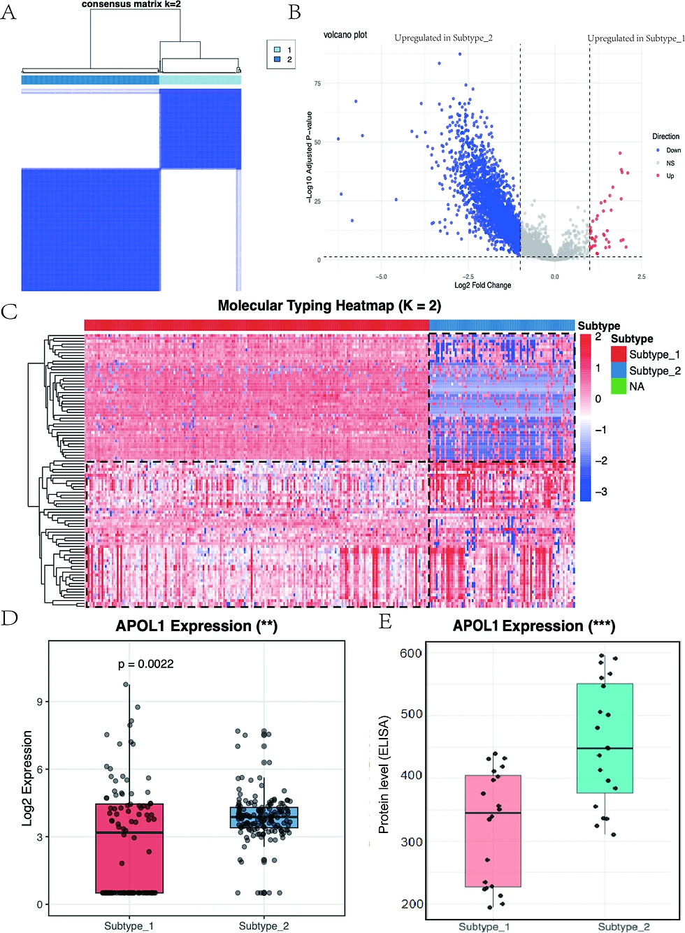
Figure 1. Two-class molecular subtyping of lupus nephritis from FFPE renal biopsy proteomics. Panel A shows the consensus clustering matrix at K=2. Panel B is a volcano plot of differential protein expression between subtypes. Panel C presents the unsupervised typing heatmap of scaled expression, annotated by subtype. Panels D and E give APOL1 boxplots, as log2 tissue expression and as serum concentration by ELISA. Adapted from Wang et al. (2026), Lupus Science & Medicine.
Where the Two Subtypes Diverge
Subtype_2 reads as the immunologically active arm. Interferon-driven innate immunity, complement activation and lysosomal handling of engulfed material surfaced together, fitting the prevailing model of how immune complexes damage the glomerulus. Subtype_1 looked structural instead, weighted toward keratinisation and epithelial remodeling. That second signature deserves scrutiny: in whole-section FFPE digests, keratin peptides can reflect specimen handling as readily as epithelial biology, and the authors do not fully separate the two.
From Subtypes to Patient Stratification
Subtype assignment moved with creatinine, eGFR and chronicity index, none of which are encoded in the ISN/RPS class. APOL1 is the most portable result, since a tissue-derived discriminator that survives transfer to a serum immunoassay can actually enter a clinic workflow. The five-protein class panel is weaker than it looks: sensitivity of 0.93 against specificity of 0.54 rules out proliferative disease reasonably well and rules it in poorly. The same stratification logic sits behind work on urinary protein signals in acute kidney injury. No longitudinal outcome data were available, so prognostic claims stay unproven.
In Practice: Running Archival Tissue at Cohort Scale
FFPE is unforgiving because crosslinking efficiency and block age both drift, and that drift imitates biology unless peptide load and batch correction across plates are locked down first. We run this class of archival-tissue DIA workflow in house at Dalton, and a cohort of this size is a good argument for fixing the gradient and the on-column amount before a single sample is queued. Where one protein layer stalls, adding transcript or metabolite readouts from the same block is usually what turns a subtype label into a decision, which is the reasoning behind our approach to multi-omics biomarker discovery and the tissue and biofluid protein profiling workflows that feed it.
Frequently Asked Questions
What is proteomics used for in clinical research?
Proteomics measures which proteins are present in a sample and how their abundance shifts with disease or treatment. Clinical studies use it to find diagnostic and stratification markers and to nominate drug targets. DIA mass spectrometry quantifies several thousand proteins per sample without a pre-selected antibody panel.
Can proteomics be run on archived FFPE biopsy samples?
Yes. Formalin crosslinks are reversed during extraction, and current DIA methods recover several thousand proteins from one biopsy section. Success depends on consistent block handling, controlled peptide load and batch correction, since variation in any of those can masquerade as a biological subtype.
What did APOL1 reveal about lupus nephritis subtypes?
APOL1 levels differed significantly between the two tissue-defined subtypes, and the same direction appeared in serum by ELISA. That dual readout makes it a candidate blood marker for assigning subtype without a repeat biopsy. The validation set was only 40 patients, so a larger cohort is needed before this means anything clinically.
Conclusion
What is believable here is that lupus nephritis carries molecular structure the histologic classes don't capture, and that archival tissue is a legitimate substrate for finding it. What isn't proven is whether either subtype predicts anything forward in time; without follow-up data, this stays descriptive. For a biomarker program, the practical read is that pairing a tissue discovery arm with a serum assay early, as the APOL1 work did, saves a translation step later.
Related Reading
See how we run these analyses in one lab: Dalton's multi-omics CRO services.
Citation
Wang, L., Wang, W., Hao, Y., Chen, L., Qi, C., Liang, D., Guo, L., Zhao, J., Sun, X., Qin, S., & Cao, L. (2026). DIA proteomics of FFPE renal biopsies reveals two molecular subtypes of lupus nephritis and identifies APOL1 as candidate biomarker for stratification. Lupus Science & Medicine, 13(2), e002123. https://doi.org/10.1136/lupus-2026-002123
Note
This blog post summarizes findings from the above-cited research. Figures are adapted from the original publication. For full details, please refer to the source article.
By Seungjun Yeo, CEO at Dalton Bioanalytics. Specializing in multi-omics mass spectrometry for drug discovery and biomarker research.